 We often don?t give much thought to the forces around us ? gravity, electromagnetic fields, sound waves, light, radio waves, radiation.?But the discovery and study of these natural forces, and our own creativity, led to the development of one of the most important fields of medicine today:
Diagnostic Imaging
Most of us know someone, or may be someone, who has had at least one diagnostic imaging study: an MRI, CT Scan, X-ray, Ultrasound, or Nuclear Medicine study. At Lexington Diagnostic Center & Open MRI, we know how important our imaging services are to our patients and their doctors. But how did we arrive at today?s use of modern medical technology? You might be surprised!
X-ray
Discovered in 1895 by Wilhelm Roentgen of W?rzburg, Germany, we started to use x-rays in the U.S. within just a few months. By the outbreak of World War I in 1914, most hospitals were equipped with x-ray machines. The ability to locate bullet fragments in soldiers became a crucial tool for saving their lives.
The general x-ray is the workhorse of diagnostic imaging and are frequently used to diagnose broken bones and other musculoskeletal conditions. Although x-rays pass through the soft tissues of the body rather easily, they are also useful in diagnosing conditions of internal organs, including the lungs (pneumonia, TB, cancer), breasts (cancer), abdomen (digestive disorders), heart (enlargement) and pelvis (reproductive disorders). A general diagnostic x-ray can help your physician make an informed decision about additional imaging or testing to aid in a diagnosis. Ultrasound Way back in the 1790?s, Italian biologist Lazzaro Spallanzani?s first studied ultrasound physics when he discovered that bats use soundwaves to navigate in the dark (echolocation). It took some time, but the idea to measure soundwaves eventually led to the development of the medical ultrasound, first used in 1956.
The principle behind ultrasound is pretty simple ? specialized equipment detects changes in high-frequency soundwaves as they pass through the body and are reflected back. A picture is built based on those changes.
Ultrasound can also capture the actual functioning of organs in real time. For example, a cardiac ultrasound allows the physician to see blood passing through the chambers of the heart, to measure the force of the blood as it leaves the heart, and to look at the heart?s valves as they open and close. Your doctor might order a cardiac ultrasound if you have symptoms such as shortness of breath, chest pains, or a murmur.
Ultrasound is the second-most commonly used diagnostic imaging procedure. It is useful for diagnosing problems of the urinary tract, gall bladder, kidney, liver, ovaries, pancreas, spleen, thyroid, uterus and blood vessels. A carotid ultrasound (and Doppler) can help physicians diagnose blocked arteries before a stroke occurs. Ultrasound involves no radiation exposure?Nuclear Medicine
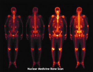
When we think ?nuclear?, our minds may go to weapons or power plants. In fact, nuclear medicine is an important and unique tool in the diagnosis and treatment of many diseases. The development of nuclear medicine spans decades and includes contributions from scientists, physicists, and engineers.
Nuclear medicine uses small amounts of radioactive substances ? or ?radiotracers? ? to get a picture of a particular area of the body. For example, radioactive iodine-125 tends to go to the thyroid gland. Measuring the thyroid?s ?uptake? of this radioactive substance allows doctors to see how the gland is functioning. In addition to the thyroid, nuclear medicine can be used to study conditions of the bones, heart, lungs, liver, and many other internal organs.
Nuclear medicine studies involve an injection or an oral dose of radiation and then imaging collects the resulting data. These procedures are extremely safe and very simple. Nuclear medicine studies measure the function of the organ or structure and, because of this, often shows abnormalities much earlier than other forms of imaging.
CT Scan (AKA the ?Cat Scan?)
The first clinical CT scan was performed on October 1, 1971 on a young lady in London, England who doctors suspected of having a brain tumor. The scan took hours to complete, but launched a new era of modern neuroimaging. Short for Computerized Axial Tomography, today?s CT scans use computer equipment and x-ray technology to create incredibly detailed, three-dimensional images quickly and comfortably. CT scans are often used in emergencies because the scan takes less than five minutes, compared to up to 30 minutes for an MRI.
The use of CT scans is on the rise, possibly because the public is increasingly aware of the signs of a stroke. Because patients are more knowledgeable than before, they?re arriving at the ER earlier in the stroke scenario, when the most can be done for them. As a result, more CT scans are being ordered so physicians can start the right treatment quickly.
CTs can also be ordered to diagnose back and spine problems, brain tumors and joint disease. An imaging procedure, called the Low Dose CT (LDCT) has recently been approved by Medicare/Medicaid as a tool for the early detection of lung cancer. This is a very promising step forward in turning the tide on this cancer. A CT scan is a powerful tool as it provides a wealth of information. However, a CT scan does expose the patient to relatively higher doses of radiation, so it?s important for you and your doctor to balance risks with benefits.
Magnetic Resonance Imaging, or MRI
When Dr. Raymond Damadian was a boy growing up in New York, his grandmother lost her painful battle with breast cancer, fueling his pursuit of a career in medical research. Dr. Damadian would eventually invent the MRI machine, which was first used to perform a full-body scan in 1977.
MRI works by using a very powerful magnet to align the hydrogen atoms inside the body. Radio waves are then used to disturb this alignment, causing the atoms to vibrate. Highly advanced computer programs generate detailed images from the vibrations. Like CT scans, detailed, three-dimensional views are available with an MRI. Unlike CT, there is no radiation exposure.
MRIs are used to diagnose a variety of disorders, including tumors, aneurysms, spinal cord injuries, multiple sclerosis, eye and inner ear problems. A form of MRI, called fMRI (functional MRI), can measure brain function. For example a head MRI can help determine whether you sustained any damage from a head injury. Your doctor may order a head MRI to investigate symptoms such as dizziness, weakness, seizures, changes in thinking or behavior, blurry vision, or chronic headaches.
Lexington Diagnostic Center and Open MRI has three MRI machines:
? Open. This equipment is very comfortable and open on 3 sides. It is an excellent option for patients who may be claustrophobic, and it can accommodate various body types.
? 1.5T. This fast-acting machine is open at both ends and provides high-quality images.
? 3T. The 3T is also open at both ends, offers very high resolution, and is excellent for orthopedic, neuro and prostate imaging.
Many brilliant minds and historical events lead to today?s options for life-saving imaging. Discuss the options with your doctor next time you need a scan, and remember that you have a choice of where to have imaging performed.? Lexington Diagnostic Center & Open MRI provides quality, state-of-the-art diagnostic imaging. We offer affordable MRI, CT, ultrasound, DEXA/Bone Density Scans, x-ray and imaged guided fluoroscopy epidural and joint injections.