Take a look inside: Nuclear Medicine Studies
When we think radioactivity, our minds naturally go to nuclear weapons or power plants. We all know radiation is DANGEROUS. But we rarely think about its beneficial uses, especially when it comes to medicine.
But radioactivity is the energy behind one of the most useful diagnostic imaging modalities in use today: nuclear medicine. The modality is unlike any other medical imaging technique because it allows the physician to see how organs, tissues and bones function at the cellular level. Even the most powerful MRI cannot do that.
How does it work?
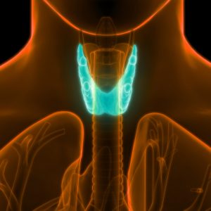
Radioactive isotopes called tracers are introduced into the body either through an injection or orally. These tracers make their way to the part of the body being studied. Interestingly, there are about 20 different radioactive isotopes that can be used and each of these tends to be attracted to certain body systems. For example, iodine-125 tends to go to the thyroid gland, making it very helpful in diagnosing hyper- and hypothyroidism.
As the tracer makes its way to the system being studied, a special camera that measures gamma radiation, records how the system processes (uptakes) the tracer. Based on these studies, the physician can determine whether the organ or system is functioning normally on a cellular level.
Nuclear medicine is useful in diagnosing a wide array of conditions affecting the bones, heart, lungs, liver and many other internal organs.
Is it safe?
Despite a 70-year track record as a safe diagnostic tool, patients sometimes express concern about a nuclear medicine study. It’s not uncommon for patients to ask jokingly if they will ‘glow’ after the exam. The short answer: No.
The amount of radiation the patient receives during a nuclear medicine study is typically quite low: usually less than that received during a routine x-ray and significantly less than that received during a CT scan.
There are three primary reasons for this. First, only a minute amount of tracer is required to capture nuclear medicine images. Tracers degrade quickly. And the body excretes them naturally, usually within 24 hours. Patients are able to resume their normal daily activities immediately following a nuclear medicine study with few restrictions. Drinking plenty of fluids will help to flush the tracer from the body.
What is nuclear medicine used to diagnose?
We’ve already discussed its use in diagnosing thyroid conditions. Here are some other ways nuclear medicine studies can help your physician:
Heart
– visualize heart blood flow and function
– detect coronary artery disease
– assess damage to the heart following a heart attack
– evaluate the results of revascularization procedures
– detect heart transplant rejection
– evaluate heart function before and after chemotherapy
Bones
– evaluate bones for fractures, infection and arthritis
– evaluate for metastatic bone disease
– evaluate painful prosthetic joints
– evaluate bone tumors
– identify sites for biopsy
Brain
– investigate abnormalities in the brain in patients with seizures, memory loss and suspected abnormalities in blood flow
– detect the early onset of neurological disorders such as Alzheimer’s disease
– assist in surgical planning and localize seizure foci
– evaluate for abnormalities in a chemical in the brain involved in controlling movement in patients with suspected Parkinson’s disease or related movement disorders
– evaluation for suspected brain tumor recurrence, surgical or radiation planning or localization for biopsy
Because nuclear medicine scans show function, they are very good at finding problems early in the course of a disease. Other modalities can determine the extent of the problem after physical change has occurred, but they cannot capture the damage in process.
If your physician or provider orders a nuclear medicine study for you don’t worry. You won’t glow and it won’t hurt. But it will provide incredibly valuable information to your doctor that he or she can use to properly diagnose and treat your condition.
The experts at Lexington Diagnostic Center and Open MRI performs a wide variety of nuclear medicine studies, including bone scans, liver/spleen studies, renal scans, bone marrow imaging, white blood cell imaging, gastric emptying, thyroid/parathyroid, and tumor scans.
For more information about nuclear medicine studies at
Lexington Diagnostic Center and Open MRI, call the office at
(859) 278-SCAN (7226).