‘Tis The Season For Rejoicing And Reflection

“Christmas is a season not only of rejoicing but of reflection.” – Winston Churchill

This is truly the most wonderful time of the year, isn’t it? As a locally owned business, the joy of the holiday season is something extra special at Lexington Diagnostic Center as we reflect on the past year and rejoice in thankfulness for our staff, patients, providers, and friends. We are continuously thankful for the patients who choose to use us for their care; for the physicians and other healthcare providers who have placed their trust and confidence in us to care for their patients; and for every member of our team who works diligently to deliver the best care to our patients and the highest quality imaging.

One of the joys of the holiday season is the opportunity to say thank you. In the spirit of the holidays, we wanted to spread some joy and share with you a few of the comments we have received from our patients this year about their experiences at LDC. We know the best way for people to get to know and trust us is through the experience of their friends and families.


RAVES ABOUT OUR TEAM

“This was the best experience I have ever had with any radiology procedure. Your staff is exceptional in making me feel comfortable and safe.”

“All of the techs were so warm and were very empathetic about me needing extra time to get to and from the various rooms because of my disability! Thank you so much!”

“I could not have completed the MRI without the sweet, smart, and professional technician that took care of me.”

“I was very impressed with the detail and care that the staff went through to make sure I understood and was comfortable with the procedure. The procedure was explained in detail by Meagan and then again by the doctor. Overall the experience was pleasant and efficient.”

“The tech that performed my CT scan was amazing! He was kind and made sure I understood and was comfortable with everything that we were doing. Don’t lose him!”

“I am always treated well and my doctors are pleased with the quality of the results. Thank you.”
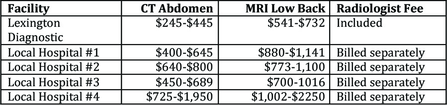
 

COMPARING PRICES

“My husband and I took our son to Lexington Diagnostic for an ultrasound. They were very accommodating to get our son in the next day and were transparent in the cost. We saved hundreds of dollars by going to LDC and we are very grateful. Thank you Lexington Diagnostic!”

“I am glad that LDC is such an affordable option and for the hours that you all are open.”

“Very friendly and accommodating staff! So many great people work there. I was in and out in no time. They saved me a ton of money, too!”

“Tech was so kind and gentle. Highly recommend and the cost savings versus a hospital setting is just mind blowing!”

Lexington Diagnostic Center & Its Employees Put Gratitude Into Action

“Gratitude can transform common days into thanksgivings, turn routine jobs into joy, and change ordinary opportunities into blessings.”  — William Arthur Ward

Lexington Diagnostic Center and Open MRI employees do more than serve patients who come to them for care. They serve the entire community, whether they ever have the opportunity to meet them, or not. “At Lexington Diagnostic Center and Open MRI, our employees embrace the fact that the most rewarding feeling is to know your actions impacted someone,” said Jesi Bowman, marketing manager.

Charles, the nuclear medicine technologist at LDC, has delivered meals for Moveable Feast once a week for the past 18 years. Moveable Feast Lexington prepares and delivers to the door a hot, nutritious meal five days a week to low-income people in Lexington-Fayette County who have HIV/AIDS or those under Hospice Care. Moveable Feast also feeds their caregivers and dependent children. Moveable Feast feeds some of the sickest and most vulnerable folks in the community. Over the 20 years since its founding, MFL has delivered more than 515,551 meals. To learn more about Moveable Feast Lexington, to donate, or to volunteer, visit their website at feastlex.org or call (859) 252-2867.

Victoria, Lexington Diagnostic Center’s front office supervisor, works with The Mack Foundation to support Lexington’s youth. The Mack Foundation is designed to promote unity, security, responsibility and versatility through a positive atmosphere for today’s youth. Programs supported by the Mack Foundation have included The Mack Mentality Mentoring program (in conjunction with the Lexington Police Athletic League); a men’s basketball tournament (a fundraiser for the Foundation); the Shelvin Mack Skills Camp; Back-to-School with Mack; and more. The Foundation emphasizes the power of positivity. “Challenges are what make life interesting. Overcoming them is what makes them meaningful.”

Paula, LDC’s Clinical Manager, currently helps pack backpacks with food for at risk children at one of the elementary schools in Fayette County. This provides food for weekends and school breaks, so they have food to eat when school is not in session. Paula can also be found serving in communities devastated by natural disasters, a facet of her mission work. This can be local such as West Liberty after the tornado in 2012, nationally, such as Mississippi after Hurricane Katrina, Houston after Hurricane Harvey, or internationally such as the earthquake in Haiti. She has trained for an early response team that waits for a call to go where needed. She and her team are expecting to go to North Carolina this fall. As an organization, Lexington Diagnostic Center also supports the student backpack program Lafayette High School.

The LDC team will be participating in the VA5K on Sunday, Nov. 4. Proceeds from the 3rd Annual Run/Walk benefit the VA Medical Center and the Lexington Fisher House, which provides lodging for families of veterans being treated at the medical center at no cost. “We love promoting healthy lifestyles and look for opportunities to get our employees active,” Bowman said.

The Center also provides support to the Kidney Health Alliance of Kentucky, whose mission it is to serve kidney disease patients and their families; increase awareness and early detection of chronic kidney disease; provide a network for collaboration among renal healthcare providers; and promote organ donation. For more information about the Kidney Alliance, visit their website at khaky.org.

The American Heart Association’s Go Red for Women campaign also receives support from Lexington Diagnostic Center. Go Red for Women seeks to raise awareness of the impact of heart disease in women. Bowman herself serves as a spokesperson for the American Heart Association in Lexington.

“Patients will always be our priority,” Bowman noted. “But service beyond scanning is integral to what we do. We put patients first, provide superior imaging services, and affordable rates. Over and above that, our employees love serving patients and their communities,” she said.

Take Action on Your Bone and Joint Health

October 2018

More than 54 percent of Americans over the age of 18 are affected by some kind of bone or joint condition, including trauma, back pain, arthritis, and osteoporosis. Three out of four people over the age of 65 suffers with a bone or joint problem.

Musculoskeletal conditions become more prevalent with age, arising from poor posture, repetitive strain, injury, and poor fitness/health habits. Bone and joint problems are not only painful, they can be disabling. In fact, musculoskeletal conditions are the most common cause of severe long-term pain and physical disability worldwide, affecting hundreds of millions of people.

Oct. 12-20 is worldwide Bone and Joint Action Week, a time to focus attention on the most common diseases affecting the bones and joints. The first step in treating bone and joint diseases is diagnosis. Often, medical imaging, in the form of CT or MRI scans is involved.

Lexington Diagnostic Center and Open MRI is the area’s only free-standing, locally owned, private diagnostic imaging center, offering state-of-the-art diagnostic imaging and therapeutic services, including:

  • Epidural steroid injections to reduce back or leg pain
  • Joint injections to alleviate pain associated with arthritis and other degenerative conditions
  • MRI and CT arthrograms to help pinpoint the cause of ongoing pain


Financial planner David Short, managing partner at Investments and Financial Planning, LLC, suffered ongoing shoulder pain off and on for more than six years. This spring the pain came back, but this time, it was severe. His primary care physician ordered an MRI arthrogram and recommended Lexington Diagnostic Center and Open MRI for his study.

During the test, musculoskeletal radiologist Jason Harris, M.D., injected a local anesthetic into the shoulder joint, followed by a special dye (contrast), that allows soft tissue, such as ligaments, cartilage, and tendons, to be imaged more readily. Short then had an MRI of his shoulder, which showed a partial tear in his labrum and damage to his rotator cuff. After a few months of physical therapy, Short is feeling good as new…and wishing he’d gotten things checked out sooner.

“At Lexington Diagnostic Center we pride ourselves on pinpointing our patients’ specific health issues, so they can receive proper treatment. We treat everyone like family and try to make the process as easy as possible,” Dr. Harris said.

For his part, Short is pleased with his experience at Lexington Diagnostic Center and recommends it to others requiring diagnostic imaging.  “They treated me very well from the first phone call, through the whole process. It was a very professional environment,” he said. “Not only was it quick and pain-free, it was affordable, as well,” he noted.

Lexington Diagnostic Center has been providing high-quality, low-cost services to area patients for 34 years. Patients choose Lexington Diagnostic Center for many reason: convenience, comfort, quality, satisfaction, and price. Lexington Diagnostic Center offers a full range of diagnostic imaging services: MRI, CT Scan, nuclear medicine, ultrasound, DEXA Scan, and general x-ray testing.

If your doctor orders imaging studies for you, be sure to call Lexington Diagnostic Center first at (859) 278-7226 to find out how they can help you get back to health – and doing the things you love – as quickly as possible.

Why do I have to undress and put on a gown? I am not wearing any metal

Lexington Diagnostic Center takes patient care and safety very seriously. If you’re familiar with imaging centers or scans, such as MRI and CT, then you’re likely aware of the fact that prior to any of these scans, a radiology technologist will politely ask you to please remove your jewelry and clothing and change into a specified gown. We ask our patients to do this to prevent injury.

Why is the gown necessary even if you’re not wearing metal? Because many different types of clothing have metal microfibers (used to help prevent shrinking) that may result in skin burns caused during the imaging scan.

The magnet on the MRI is very strong. For your safety, it is LDC’s policy that all patients undress and put on a gown to ensure that we do not get any artifacts from threads or hidden metal in your clothing. Not only for your safety, but we also want to make sure nothing obscures the images.

Now that you’re more informed, we hope you understand that if a radiologist or tech asks you to change, it’s because they’re looking out for you. To learn more about imaging scans, how to prepare for an imaging scan, what to expect during an imaging scan, or to book an imaging scan, contact us today!

859-278-7226
https://lexingtondiagnostic.com/

Lexington Diagnostic Center: Preferred By Patients & Insurers Alike

A major healthcare insurance company announced last fall that it would no longer pay for outpatient MRIs and CT scans performed at hospitals when the patient could have received the exam at a lower cost at a free-standing imaging center. So far, 13 states are affected, including Kentucky.

As you can imagine, this announcement sent shock waves through the healthcare world, especially hospitals!

Why would an insurance company implement such a policy?

The answer is simple really: cost. Hospitals carry a lot of overhead: 24-hour emergency departments, laundry and nutrition services, administration and so on. These expenses have to be made up somewhere and imaging studies are typically one source of revenue. As a result, imaging studies performed in hospitals are typically two or three times more expensive than those performed at free-standing centers.

Now if you have health insurance you might be thinking none of this matters to you: your insurance will pay for your testing. Maybe. Maybe not. It would be in your best interest to check before having an outpatient CT or MRI done to ensure that it’s being covered wherever you’re having it done.

Out of pocket expenses such as (deductibles, coinsurance, and copays) could be significantly lower than hospital costs.

Can the hospital where you’re planning to have this testing done tell you up front what your financial responsibility will be? Probably not. In all likelihood, they don’t even know.

As the area’s only free-standing, locally owned, private imaging center, Lexington Diagnostic Center has been providing high-quality, low-cost services to area patients for more than 30 years. Not only do patients experience a comfortable, home-like environment at Lexington Diagnostic Center, they enjoy peace of mind knowing an estimate of what their testing will cost up front.

Don’t believe it? Here are some examples of the costs Lexington Diagnostic Center patients have reported to us (based on Anthem BC/BS and Humana plans):

Summer Sports Injuries – Bubble Wrap, Anyone?

Warmer weather, bluer skies and sunny days have a way of spurring even the most dedicated couch potato off the cushion and into the great outdoors. Sadly, human beings don’t often behave in rational ways, we don’t like to prepare, and we often imagine ourselves to be in far better physical condition than we are.

The almost inevitable result of this behavior? Injury.

That’s not to say we shouldn’t avail ourselves of every opportunity to be more active (we should) or we shouldn’t enjoy the great outdoors (we must) or that we need to be wrapped in bubble wrap (well? maybe). But it does mean we need to be a little more careful with our bodies and to pay close attention when they’re trying to tell us something. Like “I HURT!”

The specialists at Lexington Diagnostic Center have seen it all, literally. Blown ACLs. Broken legs. Mangled elbows. Stretched ligaments. Torn cartilage. You name it, they’ve helped diagnose it. If your pursuits land you in a world of hurt this summer, the folks at Lexington Diagnostic Center can help!

In the meantime, they offer the following digest of common summer sports injuries and how they’re diagnosed to help your body survive whatever you put it through this year.

One of the most commonly occurring summer injuries is the sprained ankle. They happen everywhere: stepping down from a curb, playing badminton at the family reunion, picnicking in the park. Wherever there’s an uneven surface, there’s an opportunity for an ankle sprain! If you can put weight on the ankle for about three steps, and nothing is sticking out or looking really nasty, treatment with rest, ice, compression and elevation (RICE) should be sufficient. But if the ankle swells, becomes extremely painful or it’s difficult to walk, it’s best to see a doctor. Diagnosis will undoubtedly involve an x-ray to determine if anything’s broken.

Injuries to the shoulder can be quite painful and serious. Shoulder strain occurs when a muscle or tendon in the shoulder is stretched. Signs include pain, swelling, muscle cramps or spasms and limited range of motion. Often, these injuries occur when the weekend athlete pitches one too many fast balls, plays too much golf or tennis, or really gets at the rowing machine. A rotator cuff injury is more serious and often seen in athletes and others who use their arms overhead a lot. Think construction workers, swimmers, baseball players and weight lifters. Diagnosing shoulder injuries may include x-rays or an MRI.

Overuse injuries/stress fractures occur as people transition from an indoor running environment to pounding the pavement outside. Although increased mileage and harder surfaces are often the culprit, we also see patients with stress fractures arising out of a 5K fun run. Stress fractures aren’t fun but helping others does help lessen the pain a little (as would a little bit of training beforehand). Stress fractures can be difficult to diagnose and don’t always show up on a general x-ray. Often, an MRI is required to make the diagnosis. With a stress fracture, you’ll be on crutches (or in a boot) for a while and may be given anti-inflammatories for the swelling and pain.

Collar bone (clavicle) injuries are common among children and young adults who spend time rollerblading or at the skate park. We also see these sometimes with watersports like tubing, whitewater rafting, wakeboarding or skiing. There’s often a lot of pain and moving the arm can be very difficult. Because the collar bone lies so close to the surface of the skin, a break is usually pretty obvious, but an x-ray will confirm the diagnosis.

Knee pain is common year-round, but athletes (and would-be athletes) sometimes experience it as a result of a bike accident or from pushing themselves too far and too hard cycling. Just as in running, bicyclists should build up to longer distances over a period of weeks. If your doctor suspects a tear or injury to the meniscus (the cartilage that cushions the joint), an MRI will be ordered. If a dislocation is suspected, the first step will likely be an x-ray, possibly followed. Treatment will depend upon the severity of the injury.


Ultrasound: The Stethoscope of Modern Medicine

When you hear the word ultrasound, what springs to mind? If you’re like most, it’s probably the joy of a new life, the somewhat blurry images of babies yet to be born. As important, and as widespread, as fetal ultrasound is, there’s so much more to it than that!

Mom-to-be ultrasounds aside, this imaging technology is the second- most commonly used diagnostic imaging procedure in the United States. It has applications in diagnosing and treating heart disease; in understanding conditions affecting the circulatory system; determining the nature of various lumps and bumps; helping physicians deliver medications directly to injured areas; breaking up kidney stones; and more.

Physicians and healthcare providers turn to ultrasound so often because of its utility; its safety; the fact that patients are not exposed to radiation; its ability to record the functioning of organs in real time; its portability; and its affordability. Ultrasound technology is so useful that it has been labeled the stethoscope of modern medicine!

Principles and history of ultrasound?

As the name implies, ultrasound makes use of sound waves at very high frequencies. The principle is very similar to the way whales, dolphins, bats and other animals locate objects and navigate through their environments.

During an ultrasound, high-frequency sound waves (1 – 10 megahertz) are directed at the body structure using a hand-held device called a transducer. As sound waves bounce back, the transducer picks up the echoes and feeds data regarding their speed and intensity into a computer. The computer analyzes this data to create the ultrasound image.

Although the ancient Greeks realized the power of sound, it wasn’t until the early 20th Century that knowledge and need converge to create this powerful technique. Blast shocks from the bombing of Britain during World War II often resulted in damage to internal organs. The problem was so vexing that Royal Army Surgeon John J. Wild began searching for a non-invasive way to evaluate these injuries. Wild’s attention turned to ultrasound, which up to that time had been used in military and industrial applications.

After the war, Wild emigrated to the U.S., where he continued his work. In 1949, he discovered that sonic energy was reflected as echoes from soft biological tissues. It was an accidental discovery, but one that earned Wild recognition as the father of medical ultrasound.

Ultrasound today

The tremendous versatility and portability of ultrasound technology has made it an extremely valuable testing modality. Today, ultrasound machines can be found in the battlefield, helping to evaluate injuries and trauma and speeding appropriate treatment for military personnel.
Closer to home, Lexington Diagnostic Center provides this imaging technology in its comfortable and convenient center on Harrodsburg Road. Here, ultrasound is used to evaluate gallstones; kidney tumors; kidney stones; liver cysts and other tumors; the thyroid; blood clots in the legs; testicular masses; and abnormalities in the ovaries and uterus. Ultrasound is also used to evaluate hip problems in children and in the diagnosis of circulatory conditions.

Choosing Lexington Diagnostic Center for your medical imaging is the right choice in so many ways: convenience, comfort, quality, satisfaction, and price. In addition to ultrasound services, Lexington Diagnostic Center offers MRI, CT, nuclear medicine studies, DexaScan, and general x-ray testing. When your doctor orders a diagnostic imaging exam for you, remember – you have a choice. Call Lexington Diagnostic Center at (859) 278-7226 to learn how we can help you!

You Have the Power to Reduce Your Healthcare Costs

Regardless of whether you have the best possible health insurance, a middle-of-the-road plan, or no plan at all, healthcare costs are a continuing concern for the American family. Healthcare expenses continue to rise while wages have remained stagnant. As a result, healthcare costs (insurance premiums, deductibles and co-pays), are eating up a larger portion of the family budget now than ever before.

In fact, the average family spends about 10 percent of its annual income on healthcare-related expenses; up from 6.5 percent just a decade ago.

While it may seem there’s nothing an individual or family can do about it, there is. And if enough people take these steps, we just might be able to get healthcare costs under control.

1. Talk to your doctor about the necessity of the proposed test or procedure.

Whether you are concerned about costs or not, this is always a good discussion to have with your provider. You should never have a test or procedure without being completely clear about why it’s necessary and how it will benefit you. There’s nothing wrong with asking about alternatives that may be just as effective and less expensive.

2. Review your insurance coverage.
If you have commercial or government-sponsored insurance, it’s a good idea to review your insurance coverage to determine whether the test/procedure will be covered and under what circumstances. If it’s unclear, call your insurance company or talk to your human resources department to verify coverage. Ensure that any pre-certification requirements are met. Most physician offices will take care of this for you, but it’s always a good idea to ask. More importantly, make sure all of the providers who will be involved in your care are ‘in-network.’ It’s a pretty unpleasant surprise to receive a balance bill for an out-of-network provider when you thought everything was taken care of.

3. Ask how much it will cost.
It can be extremely difficult to get a definitive answer from hospital billing departments, benefits managers and even insurance companies. Don’t trust that online cost calculator, either! Talk to a person and take notes. Keep asking until you are quoted a price. Be sure to record who told you what it would cost and when the quote was given. You may need this when bills start arriving later. The ideal situation? Work with a provider that can tell you exactly what the cost will be – and guarantee it – without hassle or uncertainty. Lexington Diagnostic Center is one such facility. LDC patients know up front what their total cost will be, with no surprises later on.

4. Don’t assume prices are the same from one provider to the next.
They aren’t. When it comes to medical imaging, for example, hospital costs are two to three times higher than those charged by a free-standing imaging center, such as Lexington Diagnostic Center. That’s because hospitals have to carry a lot of overhead for things like the cafeteria, laundry and even the Emergency Department. Because imaging is all they do, Lexington Diagnostic Center doesn’t have those expenses to pass along. At LDC, patients pay for imaging services. That’s it. Why does this matter? Even if you have a Cadillac health plan, there’s a good chance you’ll have to pay a co-insurance. Would you rather pay 20 percent of $2,000, or 20 percent of $800?

5. Ask about fees over and above the actual hospital charges.
If you’re having a lab test, an imaging study or even surgery, you’ll receive bills from more organizations than the hospital. Understanding this concept is important. When it comes to diagnostic imaging, for example, a hospital imaging department will send you its bill (the technical component) and the radiologist, who reads and interprets the exam, will send you another bill (the professional fee). When a hospital quotes their price, it is only for the technical component. The radiologist’s fee is separate. At Lexington Diagnostic Center, our price includes both the technical component and the professional fee. You never receive a bill later for the doctor who read the exam.

Lexington Diagnostic Center has been helping patients and families in Lexington and surrounding areas save on healthcare costs for more than 30 years. Medical imaging is all we do – and we do it extremely well.

Next time your doctor orders a medical imaging exam for you MRI, CT, Ultrasound, tell him or her you prefer Lexington Diagnostic Center. We are conveniently located at 1725 Harrodsburg Road, Suite 100. For more information, please give us a call at (859) 278-7226.

Take a look inside: Nuclear Medicine Studies

Nuclear medicine studies and therapy for thyroid gland are both common procedures performed at LDC.

When we think radioactivity, our minds naturally go to nuclear weapons or power plants. We all know radiation is DANGEROUS. But we rarely think about its beneficial uses, especially when it comes to medicine.

But radioactivity is the energy behind one of the most useful diagnostic imaging modalities in use today: nuclear medicine. The modality is unlike any other medical imaging technique because it allows the physician to see how organs, tissues and bones function at the cellular level. Even the most powerful MRI cannot do that.

How does it work?
Radioactive isotopes called tracers are introduced into the body either through an injection or orally. These tracers make their way to the part of the body being studied. Interestingly, there are about 20 different radioactive isotopes that can be used and each of these tends to be attracted to certain body systems. For example, iodine-125 tends to go to the thyroid gland, making it very helpful in diagnosing hyper- and hypothyroidism.

As the tracer makes its way to the system being studied, a special camera that measures gamma radiation, records how the system processes (uptakes) the tracer. Based on these studies, the physician can determine whether the organ or system is functioning normally on a cellular level.

Nuclear medicine is useful in diagnosing a wide array of conditions affecting the bones, heart, lungs, liver and many other internal organs.

Is it safe?
Despite a 70-year track record as a safe diagnostic tool, patients sometimes express concern about a nuclear medicine study. It’s not uncommon for patients to ask jokingly if they will ‘glow’ after the exam. The short answer: No.

The amount of radiation the patient receives during a nuclear medicine study is typically quite low: usually less than that received during a routine x-ray and significantly less than that received during a CT scan.

There are three primary reasons for this. First, only a minute amount of tracer is required to capture nuclear medicine images. Tracers degrade quickly. And the body excretes them naturally, usually within 24 hours. Patients are able to resume their normal daily activities immediately following a nuclear medicine study with few restrictions. Drinking plenty of fluids will help to flush the tracer from the body.

What is nuclear medicine used to diagnose?
We’ve already discussed its use in diagnosing thyroid conditions. Here are some other ways nuclear medicine studies can help your physician:

Heart
– visualize heart blood flow and function
– detect coronary artery disease
– assess damage to the heart following a heart attack
– evaluate the results of revascularization procedures
– detect heart transplant rejection
– evaluate heart function before and after chemotherapy

Bones
– evaluate bones for fractures, infection and arthritis
– evaluate for metastatic bone disease
– evaluate painful prosthetic joints
– evaluate bone tumors
– identify sites for biopsy

Brain
– investigate abnormalities in the brain in patients with seizures, memory loss and suspected abnormalities in blood flow
– detect the early onset of neurological disorders such as Alzheimer’s disease
– assist in surgical planning and localize seizure foci
– evaluate for abnormalities in a chemical in the brain involved in controlling movement in patients with suspected Parkinson’s disease or related movement disorders
– evaluation for suspected brain tumor recurrence, surgical or radiation planning or localization for biopsy

Because nuclear medicine scans show function, they are very good at finding problems early in the course of a disease. Other modalities can determine the extent of the problem after physical change has occurred, but they cannot capture the damage in process.

If your physician or provider orders a nuclear medicine study for you don’t worry. You won’t glow and it won’t hurt. But it will provide incredibly valuable information to your doctor that he or she can use to properly diagnose and treat your condition.

The experts at Lexington Diagnostic Center and Open MRI performs a wide variety of nuclear medicine studies, including bone scans, liver/spleen studies, renal scans, bone marrow imaging, white blood cell imaging, gastric emptying, thyroid/parathyroid, and tumor scans.

For more information about nuclear medicine studies at
Lexington Diagnostic Center and Open MRI, call the office at
(859) 278-SCAN (7226).

Affordable, Effective, Lung Cancer Screening & What to Know

It should come as no surprise that Kentucky leads the nation in lung cancer deaths, especially when you consider another dubious distinction the Commonwealth holds: the highest smoking rates in the nation. Having a low-dose CT lung cancer screening is the best way to detect lung cancer early.

The criteria for the screening are strict, but are based in medical research:

– be between the ages of 55 and 80 (Medicare coverage to age 77 only) and in good enough health to withstand potential treatment for cancer, should something be found.
– have a 30-year pack history of smoking. A 30-year pack history means the patient smoked about a pack a day for 30 years, or two packs a day for 15 years, and so on.
– may currently smoke, or may have quit smoking less than 15 years ago. Congrats – after 15 years as a non-smoker, former smokers are no longer considered to be high risk!

Lexington Diagnostic Center has been performing low-dose CT screenings for lung cancer for over a decade and the screening at Lexington Diagnostic Center is very affordable. Because imaging is the focus of their business, they keep costs low and pass the savings on to our patients. LDCT screening at Lexington Diagnostic Center can be hundreds of dollars less expensive than a screening performed at a local hospital.

Talk with your primary care provider about low-dose CT screening for lung cancer and let them know you want to go to Lexington Diagnostic Center for testing. You may give LDC a call at 859-278-7226, or contact their website at lexingtondiagnostic.com.

Hosted By BT Web Group