Take a look inside: Magnetic Resonance Imaging

Magnetic Resonance Imaging - MRIMagnetic Resonance Imaging ? MRI ? is one of the most important developments in the field of medicine in the past 30 years. MRIs have become a virtual workhorse, allowing physicians to locate tumors and cysts, evaluate joint damage, pinpoint the cause of back pain, and diagnose a wide variety of conditions.

MRI technology has saved patients millions of dollars in healthcare expense; shortened time to diagnosis (and thus, treatment); allowed patients to avoid exposure to radiation; and improved the quality and length of lives of people across the U.S.

How does MRI differ from other types of medical imaging?
First, MRI studies are performed using magnetic fields. This is unique in the diagnostic imaging world, where most studies (with the exception of ultrasound) use ionizing radiation to create images of the body. There is no radiation exposure during an MRI. Instead, the equipment uses a very strong magnetic field to align the spin of hydrogen protons in the cells of the body and radio waves to cause the protons to wobble. As they wobble, the emit radio waves, which are detected by sensors in the machine.

Tissues that contain more water, such as muscles or the brain, emit stronger radio signals. Tissues that contain less water, such as bones, emit weaker signals. As it turns out, this is a perfect alignment of capabilities: X-rays are great at imaging dense tissues, such as bone, and poor at imaging soft tissues, such as muscles and brain.

You?ll sometimes hear MRI machines referred to as ?3T? or ?1.5T.? These terms refer to the strength of the magnet in relationship to the magnetic field of the earth. The larger the number, the stronger the magnetic field. (The T, by the way, honors Nikola Tesla, an electrical engineer and rival of Thomas Edison. He?s best known for his advocacy of and contributions to the development of the modern alternating current (AC) electrical system).

At Lexington Diagnostic Center and Open MRI, we have three MRI units:
? Our 0.6T open MRI is great for people with varying body types and those who have claustrophobia. The unit provides quality images and is open on three sides, making it possible for people who might not otherwise be able to undergo MRI to have this important test.

? Our 1.5T high field MRI is open on both ends, provides faster scans, and high-quality images.

? The 3T unit also is open on both ends and provides excellent quality, high-
resolution images. It is excellent for orthopedic, neuro and prostate imaging.

One of the great benefits for patients with MRI technology is that family members are usually able to accompany them in the scan room without risk of radiation exposure. Patients are able to listen to their own music (or ours) and are often in and out in less than an hour, with no restrictions on the rest of their day.

Lexington Diagnostic Center and Open MRI has been serving the needs of patients in this area for more than 30 years, providing high-quality imaging studies; compassionate, patient-centered care; great value; and easy access to needed services.

?One of the most important things people need to realize about diagnostic imaging is that they can choose where they will have their testing done? said Davonna Saier, marketing director for Lexington Diagnostic Center. ?Often people think they have to go to the hospital where their doctor is for testing, but that?s not the case.?

Because medical imaging is all lexington Diagnostic Center does, the Center is able to save its patients hundreds of dollars off the price of the same exam performed at a hospital ? These days, when patients are responsible for more and more of their healthcare costs, it really pays to shop around,? Saier said.

In addition, the cost of reading and interpreting the MRI exam (the radiologist fee) is included. Lexington Diagnostic Center patients save significantly, while receiving top-notch care and quality.

The FYI On Your MRI

Open MRI System
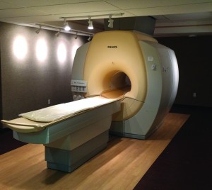
Chances are, you or someone you know has needed an MRI scan at some point. We?ve at least all heard of an MRI and might imagine?ourselves getting in to that big white tube! But what exactly are we getting ourselves in to? With the help of Paula Bracken, chief radiologic technologist at Lexington Diagnostic Center & Open MRI, let?s explore what you should know, consider, and expect when you need an MRI.

What is an MRI?
As Paula explains, ?MRI, or magnetic resonance imaging, is a non-invasive scan that uses a large magnet, pulses of radiofrequency waves, and a computer to create detailed, 2D and 3-D images of organs and structures within your body?. Physicians often use MRI imaging to diagnose conditions that may not be adequately assessed using other imaging methods such as X-ray, ultrasound or CT scan.

An MRI is often used to:
? Examine the joints, brain, spine, nerves, abdominal organs, breasts, reproductive organs and other soft tissues
? Assess blood vessels for clots and areas of narrowing
? Detect tumors and diagnose many forms of cancer
? Evaluate infections
? Assess injuries to bones, joints and muscles
? Achieve more detailed images than other imaging modalities

MRIs are often preferred over X-rays and CT scans because they don?t emit potentially harmful radiation. Here?s how it works:
1. The MRI scanner is a very strong magnet that is always turned on.
2. Since the body is made up of mostly water, hydrogen protons in the water are utilized to create an image.
3. An antenna is placed around the area of your body to be scanned.
4. You are moved into the magnetic field of the machine and the hydrogen protons go from spinning randomly to aligning with the magnetic field.
5. Radiofrequency pulses are introduced to move the protons into different positions and the antenna ?listens? to the echoes from the protons as they relax.
6. The information is sent to a computer that creates the image.

Where to have your MRI
Many patients ?go with the flow? and have their scan performed at the hospital, not realizing they have options for comfort, convenience, and cost-savings. Paula says she?s proud to provide superior care and quality at LDC. ?We offer an MRI for every need, and we take care of our patients at a fair price that they?ll find is much less expensive than at other facilities,? says Paula.

An MRI for every need
If even just the thought of that big white tube makes you cringe with claustrophobia, rest assured there?s an option for you. You can even have a loved one stay close by for support. That option is also great for little patients who want a parent close by. Lexington Diagnostic Center utilizes modern equipment and procedures to produce the best images possible while ensuring the patient?s comfort. There are three different MRI machine options to accommodate various patient needs and preferences.

In some cases, sedation may be required or requested for the patient. There is no charge for sedation at LDC. You will need to arrange for a driver to take you home.

What to expect and how to prepare:
Talk to your doctor?s referring coordinator or scheduler when your scan is ordered and they can set up your appointment with Lexington Diagnostic Center. LDC will receive your doctor?s orders and will call you to confirm your appointment. Be sure to let LDC know ahead of time if you?ve had a scan of the same area previously at another location so that images can be compared. Paula shares that LDC takes pride in making sure the entire process is easy and convenient for the patient. She says, ?We make sure patients have their appointment in a timely manner, without hassle, and are made as comfortable as possible while receiving superior image quality.?

MRI procedures are performed on an outpatient basis. Eat, drink and take your medications as usual unless instructed otherwise. If you are scheduled for an abdominal MRI, you might be asked to refrain from eating or drinking for two hours prior to your appointment.

For the scan, you?ll be asked to change into a hospital gown and remove any metal items including:
? Jewelry
? Hairpins
? Eyeglasses
? Watches
? Wigs
? Dentures
? Hearing aids

The MRI machine is a large, tube-shaped machine that the patient enters while lying comfortably on an exam table. The machine can get loud, so earplugs and headphones are available. The radiology technologist will be close by, keeping you informed and making sure you?re comfortable. You can stop the exam any time to ask questions or express concerns.

Most MRI scans take about 30 minutes to perform for each study. Afterward, the pictures will be reviewed by the radiologist and a report will be sent to your doctor.

Why choose Lexington Diagnostic Center for your MRI?
They are committed to providing the most convenient high-quality MRI imaging services for patients and their physicians. LDC offers:

? Ease of scheduling with typically same-week appointments
? Board Certified Musculoskeletal Fellowship Trained Radiologists to interpret your results
? Highly trained technologists to ensure your comfort and safety
? CD of images for each patient to have and share with their healthcare provider
? An integrated electronic medical records system that provides your referring physician easy access to your images
? Evening and Saturday appointment times
? Front door parking

PACS Portal System Offers Quality, Security, Accessibility

Lexington Diagnostic Center?s PACS Portal System Offers Quality

They were pictures of first birthdays, graduations, vacations, home renovations, the dog catching a Frisbee ? things that mattered to you in the moment. You likely shared a good number of them on Instagram or Facebook, but at least half of the photos you took were never printed or backed up.

Five years from now, you will be lucky if you can find that photo of little William destroying his first birthday cake. Even if you can, there is no guarantee the technology will be compatible.

So imagine the headache it would be if you took tens of thousands of images every year, could never delete them, and lives depended upon them being accessible a moment?s notice.

Welcome to the world of medical imaging.
Imaging studies ? X-rays, CT scans, MRIs, etc. ? are the lifeblood of modern medicine. Millions of diagnostic studies are performed in the U.S. each year, generating multiple images, each of which must be reviewed by a radiologist (the physician who specializes in interpreting medical images); permanently stored; and made accessible to the healthcare provider responsible for the patient?s care. All of this has to be available not just today, but for months and years into the future, and not just in Lexington, but anywhere the patient might seek care.

The system that makes all of this possible is called PACS ? Picture Archiving and Communications System. Since 1996, Lexington Diagnostic Center and Open MRI has been on the forefront of adopting digital imaging and using PACS to ensure imaging studies are accessible, when needed, wherever needed.

Tim Valenta, IT manager at Lexington Diagnostic Center, is responsible for ensuring all is well with the PACS system. ?Many people are relying on the images we capture at Lexington Diagnostic Center to make important decisions about treatment, and those images have to be available to them 24 hours a day, seven days a week,? Valenta said. ?It?s a responsibility I take very seriously.?

PACS is about more than just storing images and radiologists? reports, Valenta notes. Digital imaging and PACS have led to improvements in diagnosis and treatment, patient outcomes, speed, convenience and much more.

?It?s rather like the difference between a bicycle and a motorcycle,? Valenta said. Both will get you to where you need to go, but the motorcycle will get you there faster, with a lot less effort. Gone are the plates, films and chemicals needed to process the film. Gone are the dusty film storage rooms and cumbersome retrieval methods. Today, referring physicians can retrieve a patient?s study quickly over a secure Internet connection and patients leave LDC with a disc containing all of the images from their study.

Quality has improved, too. Just like the photos you take with your iPhone, sophisticated software allows the digital images to be enhanced. The radiologist can zoom in for a closer look at a particular segment of the image; change the brightness and contrast; take accurate measurements of structures; and, depending on the imaging modality, add color or create three-dimensional images.

?All of this has led to great improvements in the ability to detect changes indicative of disease or injury,? Valenta noted. Lexington Diagnostic Center began archiving its studies digitally in 1996, ensuring continuity from then to now.

The format used to store these medical images is called DICOM (Digital Imaging Communications in Medicine). The DICOM standard ensures that images captured at Lexington Diagnostic Center can be viewed at nearly any healthcare facility anywhere in the world, including a hospital in Lexington, Myrtle Beach or even London, England.

This is reassuring for patients who travel a lot or who winter in another part of the country, Valenta noted, because patients know should something happen with their health, prior imaging studies are only a few clicks away via our secure provider portal.

Accessible ? yet very secure
Keeping health information accessible to those who have a legitimate need to access it, and safe from those who do not ? is a top priority at Lexington Diagnostic Center, Valenta said.

A U.S. Navy Veteran who served as an electronics technician aboard a nuclear sub, Valenta knows about security. ?Our systems are top-notch,? he said. ?The internal network is constantly audited access restricted to only those people who need to have access to provide care to that particular patient at that particular moment.?

Referring physicians and providers can access imaging studies only through LDC?s secure provider portal and only after being granted access. Lexington Diagnostic meets both federal HIPAA and HIM guidelines, Valenta noted.

It is a big responsibility, but one that the entire staff at Lexington Diagnostic Center takes to heart. ?Every single member of our team is committed not only to providing the highest quality of imaging studies, but to protecting our patients? privacy and health information,? Valenta noted.

Terms to Know
PACS ? Picture Archiving and Communication Systems. The platform for storing and communicating digital images within a radiology practice, department or hospital.

DICOM ? Digital Imaging Communications in Medicine. The ?language? used to store and communicate medical images.

EHR/EMR ? Electronic Health/Medical Record. The ?electronic file? that contains all of a patient?s health information, including outpatient tests and treatments, office visits, hospitalizations, etc.

HIPAA ? Health Insurance Portability and Accountability Act. The federal law, passed in 1996, that both ensures that people are able to acquire health insurance as they change jobs (portability) regardless of existing medical conditions and protects the privacy of their health information. HIPAA established national standards for electronic healthcare transactions.

PHI ? Personal Health Information or Protected Health Information. The information identified by the federal government to protected from unauthorized disclosure.

HIM ? Health Information Management ? The process of acquiring, analyzing and protecting digital and traditional medical information vital to providing quality patient care.

Meet Tim Valenta
The IT manager at Lexington Diagnostic Center and Open MRI, Tim Valenta is a native of Minneapolis, Minn. He joined the U.S. Navy after graduating high school in American Fork, Utah and served six years aboard a nuclear submarine as an electronics technician.

Upon returning home, Valenta served 18 months as a missionary in Argentina. Prior to joining Lexington Diagnostic Center in 1995, Valenta worked for Unisys Corp. and with Fonar, one of the first manufacturers of MRI systems in the U.S.

Today, Valenta is responsible for designing and maintaining Lexington Diagnostic Center?s information technology network, computer systems, communications equipment, and PACS system. He also provides custom programming solutions to improve workflow at LDC. He has more than 30 years experience with electronics and computer systems.

Lexington Diagnostic Center offers High-Field MRI, Open MRI, CT, Ultrasound, X-ray, Nuclear Medicine, DEXA, and image-guided joint and epidural injections.

Hosted By BT Web Group