Take a look inside: Nuclear Medicine Studies
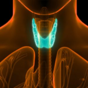
Nuclear medicine studies and therapy for thyroid gland are both common procedures performed at LDC.

When we think radioactivity, our minds naturally go to nuclear weapons or power plants. We all know radiation is DANGEROUS. But we rarely think about its beneficial uses, especially when it comes to medicine.

But radioactivity is the energy behind one of the most useful diagnostic imaging modalities in use today: nuclear medicine. The modality is unlike any other medical imaging technique because it allows the physician to see how organs, tissues and bones function at the cellular level. Even the most powerful MRI cannot do that.

How does it work?
Radioactive isotopes called tracers are introduced into the body either through an injection or orally. These tracers make their way to the part of the body being studied. Interestingly, there are about 20 different radioactive isotopes that can be used and each of these tends to be attracted to certain body systems. For example, iodine-125 tends to go to the thyroid gland, making it very helpful in diagnosing hyper- and hypothyroidism.

As the tracer makes its way to the system being studied, a special camera that measures gamma radiation, records how the system processes (uptakes) the tracer. Based on these studies, the physician can determine whether the organ or system is functioning normally on a cellular level.

Nuclear medicine is useful in diagnosing a wide array of conditions affecting the bones, heart, lungs, liver and many other internal organs.

Is it safe?
Despite a 70-year track record as a safe diagnostic tool, patients sometimes express concern about a nuclear medicine study. It’s not uncommon for patients to ask jokingly if they will ‘glow’ after the exam. The short answer: No.

The amount of radiation the patient receives during a nuclear medicine study is typically quite low: usually less than that received during a routine x-ray and significantly less than that received during a CT scan.

There are three primary reasons for this. First, only a minute amount of tracer is required to capture nuclear medicine images. Tracers degrade quickly. And the body excretes them naturally, usually within 24 hours. Patients are able to resume their normal daily activities immediately following a nuclear medicine study with few restrictions. Drinking plenty of fluids will help to flush the tracer from the body.

What is nuclear medicine used to diagnose?
We’ve already discussed its use in diagnosing thyroid conditions. Here are some other ways nuclear medicine studies can help your physician:

Heart
– visualize heart blood flow and function
– detect coronary artery disease
– assess damage to the heart following a heart attack
– evaluate the results of revascularization procedures
– detect heart transplant rejection
– evaluate heart function before and after chemotherapy

Bones
– evaluate bones for fractures, infection and arthritis
– evaluate for metastatic bone disease
– evaluate painful prosthetic joints
– evaluate bone tumors
– identify sites for biopsy

Brain
– investigate abnormalities in the brain in patients with seizures, memory loss and suspected abnormalities in blood flow
– detect the early onset of neurological disorders such as Alzheimer’s disease
– assist in surgical planning and localize seizure foci
– evaluate for abnormalities in a chemical in the brain involved in controlling movement in patients with suspected Parkinson’s disease or related movement disorders
– evaluation for suspected brain tumor recurrence, surgical or radiation planning or localization for biopsy

Because nuclear medicine scans show function, they are very good at finding problems early in the course of a disease. Other modalities can determine the extent of the problem after physical change has occurred, but they cannot capture the damage in process.

If your physician or provider orders a nuclear medicine study for you don’t worry. You won’t glow and it won’t hurt. But it will provide incredibly valuable information to your doctor that he or she can use to properly diagnose and treat your condition.

The experts at Lexington Diagnostic Center and Open MRI performs a wide variety of nuclear medicine studies, including bone scans, liver/spleen studies, renal scans, bone marrow imaging, white blood cell imaging, gastric emptying, thyroid/parathyroid, and tumor scans.

For more information about nuclear medicine studies at
Lexington Diagnostic Center and Open MRI, call the office at
(859) 278-SCAN (7226).
So Your Child Needs An MRI. Now What?

When a doctor recommends an MRI or CT scan for your child, your mind races with a million questions: Is it safe? What?s wrong?

Will it help diagnose the problem? Do they know how to take care of a child having an MRI? Will it hurt? Will she be scared? What can I do to make him feel more comfortable? What if she can?t hold still for the test? What if he freaks out? What if I freak out?

It?s normal to be concerned, but the first thing you need to do is to relax yourself! Remember, kids are like little sponges; they soak up the tension, concern and worry exuded by parents and caregivers and squeeze it out as the time for the test or procedure grows near.

Across the U.S., people of all ages undergo MRIs and other medical imaging procedures every day. Sure there are precautions that must be taken, but the healthcare professionals who will perform the tests are just that ? professionals. They have received extensive and ongoing training in caring for both children and adults, and they and put that training into practice every day!

If there?s time, and there usually is, it?s best to talk to your child about the exam and what it will entail, said Karen Sykes, a nurse at Lexington Diagnostic Center and Open MRI. Sykes works directly with children undergoing diagnostic imaging, and their parents, to ensure they have the most comfortable experience possible.

There are a variety of ways to talk to your child about an MRI or CT, Sykes said. For younger kids, parents may compare the experience to getting a photograph taken. It?s something they are all familiar with (think smart phone cameras and selfies) and so it?s no big deal.

Kids who are especially inquisitive may want to know about the kind of ?camera? being used and how it can take pictures of things inside your body. Showing them MRI and CT images online can help them understand and most kids are excited to learn that they will leave Lexington Diagnostic Center with a disk of their images they can view at home!

Older children may want to know about the science of an MRI or a CT. For them, you may want to do a little research so you can answer basic questions. ?I recommend parents do some online research,? Sykes said. ?There are videos available that show pediatric MRIs and teaches kids what will happen during the test.? Familiarity will be reassuring to children. YouTube has both live action and animated videos.

Some parents employ a little ?bribery? to ensure cooperation: a special treat after successful completion of the exam. For a little one, that might mean an ice cream cone. For an older child, perhaps it?s a Mommie-and-Me day at the beauty salon or a new video game. Make it something you?ll both look forward to and remember

Reassure your child that the people who will be taking care of them will do their very best for them. At Lexington Diagnostic Center, a parent or loved one may sit or stand beside the MRI machine so the child never feels alone in the exam room.

During the actual exam, the technologist may ask the child to play a little game that will help them to hold very still. The child may be asked to pretend they are in a rocket ship taking off for the moon; or are a statue in the park; or even frozen. The games are designed to help the child hold still during the active scan period.

Depending on the test, you may be able to accompany the child into the scan room during the procedure. Kids can choose their own music during the scan and younger ones may be able to take a favorite blanket into the scanner with them. All kids having a scan at Lexington Diagnostic Center receive a stuffed animal to take home after the test.

A lot of parents ask about open MRI for their kids, Sykes said, but this equipment may not be the best choice ? it requires children to hold still for much longer periods of time, Sykes noted. ?Parents ask about open MRI because they may be a little claustrophobic themselves,? she said, ?But it?s important to remember there?s a lot more room in an MRI machine when you?re a 50-pound child than when you are 200-pound adult male. Claustrophobia may not even be a problem.?

?The vast majority of the time, we can get a scan done for a child when no one else has been able to,? Sykes said, ?because we work very hard to help the child and parent feel comfortable and at ease. Our facility isn?t as scary as a hospital and we certainly take the time to work with them one-to-one,? she noted. ?We treat every patient with warmth and compassion and we are especially skilled at working with patients and children with special needs, including infants, children, the elderly and those with developmental disabilities.?

If your child is scheduled to have an MRI, CT scan, ultrasound, nuclear medicine or even a general X-ray, give Lexington Diagnostic Center a call at (859) 278-7226 Or call toll free 800-755-7441 to learn more about our child ? and family-friendly services. You?ll be glad you did.

Meet Nurse Karen Sykes
Karen Sykes, a licensed practical nurse, is originally from Elkhorn City deep in the mountains of Eastern Kentucky.

She returned to the mountains after attending school at Eastern Kentucky University. It was at this time that she found her life?s work: nursing. Sykes graduated from the nursing program at Mayo Tech in 1982. During her career, she has worked in all areas of nursing, including pediatrics, cardiac, geriatrics, critical care, neurology, IV therapy and emergency services.

Sykes came to Lexington Diagnostic Center in 1999 to ?help out for a couple of days? while the practice searched for a full-time nurse. After a short time, Sykes came to realize Lexington Diagnostic was where she needed to be.

Seventeen years later, Sykes provides all of the nursing care at the Center and serves on the practice?s management team. She, and her staff, will take on any challenge, but patient care is her specialty. Her patients often comment on how kind and caring she is, how they felt like they were the only patient Sykes had that day. Her compassion and experience make it possible for her to care for a diverse patient population, including infants, those with special needs, the very ill and elderly.

A mother of two (and grandmother, too), Sykes knows what it?s like to have a sick child and works hard to put parents? minds at ease and to answer all questions in a way that is complete, concise and understandable.

PACS Portal System Offers Quality, Security, Accessibility

Lexington Diagnostic Center?s PACS Portal System Offers Quality

They were pictures of first birthdays, graduations, vacations, home renovations, the dog catching a Frisbee ? things that mattered to you in the moment. You likely shared a good number of them on Instagram or Facebook, but at least half of the photos you took were never printed or backed up.

Five years from now, you will be lucky if you can find that photo of little William destroying his first birthday cake. Even if you can, there is no guarantee the technology will be compatible.

So imagine the headache it would be if you took tens of thousands of images every year, could never delete them, and lives depended upon them being accessible a moment?s notice.

Welcome to the world of medical imaging.
Imaging studies ? X-rays, CT scans, MRIs, etc. ? are the lifeblood of modern medicine. Millions of diagnostic studies are performed in the U.S. each year, generating multiple images, each of which must be reviewed by a radiologist (the physician who specializes in interpreting medical images); permanently stored; and made accessible to the healthcare provider responsible for the patient?s care. All of this has to be available not just today, but for months and years into the future, and not just in Lexington, but anywhere the patient might seek care.

The system that makes all of this possible is called PACS ? Picture Archiving and Communications System. Since 1996, Lexington Diagnostic Center and Open MRI has been on the forefront of adopting digital imaging and using PACS to ensure imaging studies are accessible, when needed, wherever needed.

Tim Valenta, IT manager at Lexington Diagnostic Center, is responsible for ensuring all is well with the PACS system. ?Many people are relying on the images we capture at Lexington Diagnostic Center to make important decisions about treatment, and those images have to be available to them 24 hours a day, seven days a week,? Valenta said. ?It?s a responsibility I take very seriously.?

PACS is about more than just storing images and radiologists? reports, Valenta notes. Digital imaging and PACS have led to improvements in diagnosis and treatment, patient outcomes, speed, convenience and much more.

?It?s rather like the difference between a bicycle and a motorcycle,? Valenta said. Both will get you to where you need to go, but the motorcycle will get you there faster, with a lot less effort. Gone are the plates, films and chemicals needed to process the film. Gone are the dusty film storage rooms and cumbersome retrieval methods. Today, referring physicians can retrieve a patient?s study quickly over a secure Internet connection and patients leave LDC with a disc containing all of the images from their study.

Quality has improved, too. Just like the photos you take with your iPhone, sophisticated software allows the digital images to be enhanced. The radiologist can zoom in for a closer look at a particular segment of the image; change the brightness and contrast; take accurate measurements of structures; and, depending on the imaging modality, add color or create three-dimensional images.

?All of this has led to great improvements in the ability to detect changes indicative of disease or injury,? Valenta noted. Lexington Diagnostic Center began archiving its studies digitally in 1996, ensuring continuity from then to now.

The format used to store these medical images is called DICOM (Digital Imaging Communications in Medicine). The DICOM standard ensures that images captured at Lexington Diagnostic Center can be viewed at nearly any healthcare facility anywhere in the world, including a hospital in Lexington, Myrtle Beach or even London, England.

This is reassuring for patients who travel a lot or who winter in another part of the country, Valenta noted, because patients know should something happen with their health, prior imaging studies are only a few clicks away via our secure provider portal.

Accessible ? yet very secure
Keeping health information accessible to those who have a legitimate need to access it, and safe from those who do not ? is a top priority at Lexington Diagnostic Center, Valenta said.

A U.S. Navy Veteran who served as an electronics technician aboard a nuclear sub, Valenta knows about security. ?Our systems are top-notch,? he said. ?The internal network is constantly audited access restricted to only those people who need to have access to provide care to that particular patient at that particular moment.?

Referring physicians and providers can access imaging studies only through LDC?s secure provider portal and only after being granted access. Lexington Diagnostic meets both federal HIPAA and HIM guidelines, Valenta noted.

It is a big responsibility, but one that the entire staff at Lexington Diagnostic Center takes to heart. ?Every single member of our team is committed not only to providing the highest quality of imaging studies, but to protecting our patients? privacy and health information,? Valenta noted.

Terms to Know
PACS ? Picture Archiving and Communication Systems. The platform for storing and communicating digital images within a radiology practice, department or hospital.

DICOM ? Digital Imaging Communications in Medicine. The ?language? used to store and communicate medical images.

EHR/EMR ? Electronic Health/Medical Record. The ?electronic file? that contains all of a patient?s health information, including outpatient tests and treatments, office visits, hospitalizations, etc.

HIPAA ? Health Insurance Portability and Accountability Act. The federal law, passed in 1996, that both ensures that people are able to acquire health insurance as they change jobs (portability) regardless of existing medical conditions and protects the privacy of their health information. HIPAA established national standards for electronic healthcare transactions.

PHI ? Personal Health Information or Protected Health Information. The information identified by the federal government to protected from unauthorized disclosure.

HIM ? Health Information Management ? The process of acquiring, analyzing and protecting digital and traditional medical information vital to providing quality patient care.

Meet Tim Valenta
The IT manager at Lexington Diagnostic Center and Open MRI, Tim Valenta is a native of Minneapolis, Minn. He joined the U.S. Navy after graduating high school in American Fork, Utah and served six years aboard a nuclear submarine as an electronics technician.

Upon returning home, Valenta served 18 months as a missionary in Argentina. Prior to joining Lexington Diagnostic Center in 1995, Valenta worked for Unisys Corp. and with Fonar, one of the first manufacturers of MRI systems in the U.S.

Today, Valenta is responsible for designing and maintaining Lexington Diagnostic Center?s information technology network, computer systems, communications equipment, and PACS system. He also provides custom programming solutions to improve workflow at LDC. He has more than 30 years experience with electronics and computer systems.

Lexington Diagnostic Center offers High-Field MRI, Open MRI, CT, Ultrasound, X-ray, Nuclear Medicine, DEXA, and image-guided joint and epidural injections.

Hosted By BT Web Group