CT Scan
CT is a non-invasive test that assists your provider in diagnosing medical conditions. CT, sometimes called a CAT scan, uses x-rays and a specialized computer to create images as if we are slicing a loaf of bread and looking at each slice individually. These images may look at the internal organs, brain, soft tissues, or bones and blood vessels. If we are looking at the internal organs in the abdomen or pelvis, you will be asked to drink barium. Barium will outline your stomach and bowels to aid in diagnosis. Your CT scan may also require an injection of contrast. This will require an IV to administer the contrast.
A CT scan only takes a few minutes to complete. Some people have described the scanner as looking like a donut.
Ionizing radiation is used for CT. Lexington Diagnostic Center is committed to using the lowest possible dose of radiation to obtain a high quality image. We have pledged to Image Gently www.imagegently.org and Image Wisely www.imagewisely.org
There is generally no prep required for a CT scan. You may be asked to remove certain articles of clothing and jewelry if these items will interfere with the images.
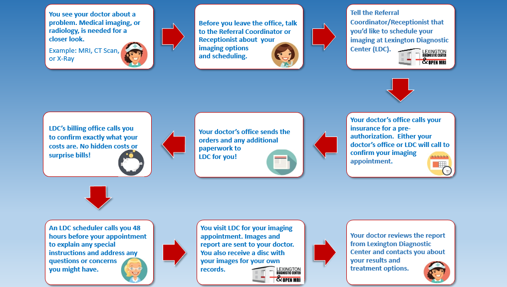
After your study is completed, the images will be burned on a CD for you to take with you. Your provider may want to view the images, so be sure to take the CD with you to your follow-up appointment. The images will also be sent to a high-resolution computer screen for the radiologist (a physician who specializes in diagnostic imaging) to interpret. Your provider will receive a written report about the results within 3 business days.
MRI
MRI is a non-invasive test that assists your provider in diagnosing medical conditions. MRIs are performed using a strong magnetic field, radiofrequency pulses and a computer to create detailed images of internal organs, the brain, spine, soft tissues, and bones and joints. An MRA uses the same scanner to image your blood vessels. An MRI or an MRA may require an injection of contrast. This will require an IV to administer the contrast.
MRI does not use any ionizing radiation, but there are other safety concerns for MRI. Since MRI uses a strong magnetic field, which can attract ferromagnetic objects, and radiofrequency pulses, that can cause heating in certain implanted devices or bullets and shrapnel, it is very important for us to know what implants or injuries you might have. You may also have something on the outside such as an insulin pump, tens unit or medication patch. We may need to do some research about whatever implant(s) you might have to determine its safety for MRI. You will be asked questions about these types of things several times before and during your visit. Please be patient we are doing this because your safety is our number one priority!
There is generally no prep required for an MRI, but you will be asked to put on a gown for your MRI. You will also need to remove jewelry, watches, hair pins and any other metallic objects. You will not be able to bring your purse, wallet, cell phone, lighter, pocketknife or other items in your pockets into the MRI room remember, the scanner is a very strong magnet! A locker will be made available so you can lock up your belongings. You also may want to consider leaving jewelry or other valuables at home.
After your study is completed, the images will be burned on a CD for you to take with you. Your provider may want to view the images, so be sure to take the CD with you to your follow-up appointment. The images will also be sent to a high-resolution computer screen for the radiologist (a physician who specializes in diagnostic imaging) to interpret. Your provider will receive a written report about the results within 3 business days.
Nuclear Medicine
Nuclear Medicine is a non-invasive test that assists your provider in diagnosing medical conditions or to treat hyperthyroidism. Nuclear Medicine is performed by using radiotracers injected through an IV or swallowing a pill. These images may look at the internal organs, soft tissues, thyroid gland, or bones. Nuclear Medicine looks at molecular activity, structure and function.
The radiotracer is radioactive. It contains small amounts of radiation and results in a low radiation dose to the patient. Nuclear Medicine has been used for over 5 decades and there are no known long term effects from the small dose of radiation you will receive.
Depending on the specific test being performed, it may take the radiotracer anywhere from seconds to several days to accumulate in the area to be imaged. You may have the test immediately after you are given the radiotracer or you may be required to return after a few hours or a few days. The technologist will give you specific instructions about when you will need to return for imaging.
There is a prep required for some scans. You will be given specific instructions prior to coming for your nuclear medicine procedure.
After your study is completed, the images will be burned on a CD for you to take with you. Your provider may want to view the images, so be sure to take the CD with you to your follow-up appointment. The images will also be sent to a high-resolution computer screen for the radiologist (a physician who specializes in diagnostic imaging) to interpret. Your provider will receive a written report about the results within 3 business days.
Ultrasound
Ultrasound is a non-invasive test that assists your provider in diagnosing medical conditions. Ultrasound, sometimes called sonography, uses high frequency sound waves through a transducer (probe) which is held against the body part being scanned. A warm gel will be ‘squirted’ between your skin and the probe. The gel allows the probe to make good contact with your skin. The technologist may need to press firmly to obtain the best pictures. You may be asked to change your position or hold your breath. These images may look at the internal organs, soft tissues, thyroid gland or blood vessels. Ultrasound sees structure and movement.
No radiation is used for ultrasound and the procedure is safe and painless.
You may be asked to remove certain articles of clothing in order to expose the skin in the area to be scanned. The technologist will respect your privacy at all times and will use a sheet or towel to minimize the exposed area. After the test, the technologist will attempt to wipe away the gel or hand you a towel to wipe it away so it does not get on your clothing.
There is a prep required for some scans. It is difficult for ultrasound waves to ‘see’ through air, so the goal of the prep is to reduce the amount of gas in your stomach and bowels. If you are scheduled for an ultrasound anywhere in the abdomen, including the kidneys or blood vessels, you should not eat, chew gum, drink carbonated beverages or smoke for 6 hours before your scan.
If the test you are scheduled for is to look at the blood vessels that feed your kidneys (renal arterial duplex), in addition to the prep above, take 2 Gas-Ex tablets the night before your test and 2 more Gas-Ex tablets 2 hours before your appointment time.
After your study is completed, the images will be burned on a CD for you to take with you. Your provider may want to view the images, so be sure to take the CD with you to your follow-up appointment. The images will also be sent to a high-resolution computer screen for the radiologist (a physician who specializes in diagnostic imaging) to interpret. Your provider will receive a written report about the results within 3 business days.
DXA
DXA (dual-energy x-ray absorptiometry) is a non-invasive test that assists your provider in diagnosing osteopenia and osteoporosis by measuring your bone mineral density. DXA may also be called bone density scanning, bone densitometry or DEXA.
There is a small amount of ionizing radiation with this test. This test is very safe.
You will lie on your back and the x-ray machine will pass over you while it takes x-rays of your spine and both hips. If it is difficult for you to lie on your back, you can sit in a chair and the DXA will x-ray the bones in your forearm.
If your provider has also ordered an LVA (lateral vertebral assessment), you will be asked to turn onto your side. The scanner will x-ray a side view of your spine to evaluate for spine fractures.
There is a prep for a DXA exam. You should not take calcium supplements for 48 hours before your test. If your provider has also ordered tests that require you to have barium (e.g. CT scan of your abdomen, GI series or barium enema), a nuclear medicine test or a test that requires an injection of IV contrast (e.g. CT scan or IVP), you should have your DXA done first or wait for 3 days after any of those tests have been completed.
After your study is completed, the computer will compare your results with other people in your age group and of the same size and gender and generate a report. The radiologist (a physician who specializes in diagnostic imaging) will analyze the images and report. Your provider will receive a written report about the results within 3 business days.
X-ray
X-ray is a non-invasive test that assists your provider in diagnosing medical conditions. X-rays are performed using a small amount of ionizing radiation to capture images of the inside of your body such as of bones, lungs, heart, abdomen, and pelvis.
There is no prep required for x-rays, but you may be asked to remove certain articles of clothing or jewelry depending on the body part to be x-rayed.
X-rays have been around longer than any other imaging modality and are the most common medical imaging test.
