Call & Compare Prices!

Recently we had a patient quoted $785 for her ultrasound at her local hospital. With us, the cost was $154.07. We also had a patient who planned to pay over $500 for a nasal bone x-ray at the hospital. She called to compare prices here and ended up seeing us because we only charged her $27.30.

We save patients hundreds of dollars every day, and the radiology reading fee is included in the price. If your healthcare provider tells you that you need an MRI, CT, x-ray, or nuclear medicine, please know you have a choice! Call and compare prices at Lexington Diagnostic Center. We would love to care for your radiology needs and your financial health.

Take a look inside: Magnetic Resonance Imaging

Magnetic Resonance Imaging - MRIMagnetic Resonance Imaging ? MRI ? is one of the most important developments in the field of medicine in the past 30 years. MRIs have become a virtual workhorse, allowing physicians to locate tumors and cysts, evaluate joint damage, pinpoint the cause of back pain, and diagnose a wide variety of conditions.

MRI technology has saved patients millions of dollars in healthcare expense; shortened time to diagnosis (and thus, treatment); allowed patients to avoid exposure to radiation; and improved the quality and length of lives of people across the U.S.

How does MRI differ from other types of medical imaging?
First, MRI studies are performed using magnetic fields. This is unique in the diagnostic imaging world, where most studies (with the exception of ultrasound) use ionizing radiation to create images of the body. There is no radiation exposure during an MRI. Instead, the equipment uses a very strong magnetic field to align the spin of hydrogen protons in the cells of the body and radio waves to cause the protons to wobble. As they wobble, the emit radio waves, which are detected by sensors in the machine.

Tissues that contain more water, such as muscles or the brain, emit stronger radio signals. Tissues that contain less water, such as bones, emit weaker signals. As it turns out, this is a perfect alignment of capabilities: X-rays are great at imaging dense tissues, such as bone, and poor at imaging soft tissues, such as muscles and brain.

You?ll sometimes hear MRI machines referred to as ?3T? or ?1.5T.? These terms refer to the strength of the magnet in relationship to the magnetic field of the earth. The larger the number, the stronger the magnetic field. (The T, by the way, honors Nikola Tesla, an electrical engineer and rival of Thomas Edison. He?s best known for his advocacy of and contributions to the development of the modern alternating current (AC) electrical system).

At Lexington Diagnostic Center and Open MRI, we have three MRI units:
? Our 0.6T open MRI is great for people with varying body types and those who have claustrophobia. The unit provides quality images and is open on three sides, making it possible for people who might not otherwise be able to undergo MRI to have this important test.

? Our 1.5T high field MRI is open on both ends, provides faster scans, and high-quality images.

? The 3T unit also is open on both ends and provides excellent quality, high-
resolution images. It is excellent for orthopedic, neuro and prostate imaging.

One of the great benefits for patients with MRI technology is that family members are usually able to accompany them in the scan room without risk of radiation exposure. Patients are able to listen to their own music (or ours) and are often in and out in less than an hour, with no restrictions on the rest of their day.

Lexington Diagnostic Center and Open MRI has been serving the needs of patients in this area for more than 30 years, providing high-quality imaging studies; compassionate, patient-centered care; great value; and easy access to needed services.

?One of the most important things people need to realize about diagnostic imaging is that they can choose where they will have their testing done? said Davonna Saier, marketing director for Lexington Diagnostic Center. ?Often people think they have to go to the hospital where their doctor is for testing, but that?s not the case.?

Because medical imaging is all lexington Diagnostic Center does, the Center is able to save its patients hundreds of dollars off the price of the same exam performed at a hospital ? These days, when patients are responsible for more and more of their healthcare costs, it really pays to shop around,? Saier said.

In addition, the cost of reading and interpreting the MRI exam (the radiologist fee) is included. Lexington Diagnostic Center patients save significantly, while receiving top-notch care and quality.

Understanding Diagnostic Imaging: The CT Scan

Lightspeed 16 Understanding Diagnostic Imaging The CT Scan
Today, the ease with which modern medicine diagnoses illnesses and injuries is almost taken for granted. It seems as though there?s almost nothing that cannot be discovered, diagnosed and treated.

It wasn?t always that way.

One of the most important developments in modern medicine was harnessing the power of X-rays to peer inside the human body. Before 1895, when X-rays were first used in medicine, physicians relied almost entirely upon the physical exam, their own knowledge and a little bit of luck to diagnose broken bones, tumors and even gunshot wounds.

Today, physicians still use their knowledge and expertise ? but those aren?t the only tools available to them. Modern imaging techniques allow physicians to see inside the brain, lungs, heart, abdomen, joints, spine ? just about every body structure. We have technology not just to see organs, but to see them at work.

One of the most powerful tools in the medical imaging arsenal is computed axial tomography, also known as CAT scan or CT scan. The CT scan is a relatively new technology, that uses X-rays along with computer technology to produce high-definition, detailed images of the soft tissues of the body. Its primary uses are in imaging the brain, lungs, pulmonary arteries, spine, and joints.

British engineer Godfrey Hounsfield and South African physicist Allan Cormack invented CT scan technology, in 1972, with a research budget of just $40,000. The very first scan took nine days to complete and used gamma radiation. The scan involved 160 passes around the object, one degree at a time. The computed part of the scan took several additional days to complete.

By using x-rays rather than gamma rays, the pair were able to reduce scan times significantly (to nine hours). By 1974, the technology was ready for medical application. These first scanners could only be used on the head and brain. In 1976, CT scanners were able to scan larger body parts, including the chest.

Fortunately, scanning and computer technology have improved vastly since CT technology was first introduced. Today, a chest CT takes just a matter of seconds, and results are available almost immediately.

Hounsfield and Cormack won the Nobel Prize for Medicine and Physiology with their medical innovation. Just as importantly, physicians gained access to a powerful new tool that has dramatically improved the ability to diagnose, treat and monitor disease. There are about 6,000 CT scanners in the U.S. today, with more than 72 million CTs performed annually.

CT scanning technology has helped patients by reducing uncertainty, trial-by-error and the need for exploratory surgery. One of the most exciting developments in CT scan technology is its use as a screening tool for lung cancer. It is the first, and only, screening that has been recognized by the U.S. Preventative Services Task Force as highly effective. Lexington Diagnostic Center has led the region in offering the service for more than 10 years.

?CT scanning technology really revolutionized medicine,? said radiologist Jason Harris, M.D., medical director/owner of Lexington Diagnostic Center. ?But as with everything we do in medicine, there are potential risks,? he noted. Among these is that CT scans expose patients to ionizing radiation, roughly equivalent to that of 200 chest x-rays. ?As powerful as this technology is, a patient should have a CT scan only when it is really necessary,? he noted.

To further ensure safety, patients should verify that any facility where they might receive testing follows the strictest protocols for safety and is accredited by an independent accreditation organization. Accreditation is the public?s assurance that the center meets or exceeds accepted safety standards including ALARA standards (As Low As Reasonably Achievable). Lexington Diagnostic Center is accredited by the Intersocietal Accreditation Commission for CT services.

Affordable, Effective Lung Cancer Screening at Lexington Diagnostic Center

Affordable, Effective Lung Cancer Screening at Lexington Diagnostic CenterIt should come as no surprise that Kentucky leads the nation in lung cancer deaths, especially when you consider another dubious distinction the Commonwealth holds: the highest smoking rates in the nation.

Because smoking is the No. 1 cause of lung cancer, the single most important thing you can do to reduce your risk of developing lung cancer is to stop smoking. The minute you stop smoking, your body begins to heal and your risk starts to fall. Within two months of stopping, your lung function begins to improve; after 10 years, your risk of being diagnosed with lung cancer is 30 to 50 percent of that of a smoker.

Regardless of whether you?ve recently quit smoking, you quit five years ago, or you continue to smoke, as they age, most people begin to think about their lungs and the damage they?ve inflicted by smoking. Unfortunately, there hasn?t always been a good way to detect lung cancer early, when the chances for a cure are greatest.

A few years ago, the U.S. Preventive Services Task Force (USPSTF) came out in support of a screening exam that a large-scale study demonstrated was effective at finding lung cancer early. The test is called low-dose computed tomography, or LDCT. At Lexington Diagnostic Center and Open MRI, we?ve been offering this testing for over a decade, even before the USPSTF published its findings.

Along with the USPSTF support came a recommendation that people at high risk for developing lung cancer receive the screening every year. As a result, private insurers (such as Anthem, Cigna and United Healthcare) may cover the cost of the exam for eligible patients, as do Medicare and Medicaid.

Who?s eligible for the screening
The criteria for the screening are strict, but are based in medical research. To be eligible for the screening:

  • The individual must be between the ages of 55 and 80 (Medicare coverage to age 77 only) and in good enough health to withstand potential treatment for cancer, should something be found.
  • The person must have a 30-year pack history of smoking. A 30-year pack history means the patient smoked about a pack a day for 30 years, or two packs a day for 15 years, and so on.
  • The individual may currently smoke, or may have quit smoking less than 15 years ago. Congrats ? after 15 years as a non-smoker, former smokers are no longer considered to be high risk!
  • Annual screening is to be discontinued when the patient has been smoke-free for 15 years or if their health declines to a level at which they would no longer be able (or interested) in curative treatment for lung cancer.


It may seem odd that people who have symptoms of lung cancer are not eligible for the screening. But remember, the screening is designed to detect cancer early. If you have symptoms, by definition, early detection is off the table and a different set of protocols applies.

What does the screening involve?
The screening is quick, simple and painless. Patients are taken to a private room, where they may be asked to change into an exam gown. They are then positioned on a table and the technologist leaves the room. The technologist will be in constant contact with the patient throughout the exam. The table will move into the CT tube and a series of X-rays will be taken. Patients may be asked to hold their breath for short periods of time. When the exam is finished, the table will move out of the tube and the technologist will assist the patient from the table. The actual exam takes no more than 10 minutes.

The images will be read by LDC?s radiologist and the findings sent to the referring physician within two business days.

The Lexington Diagnostic Center advantage
Lexington Diagnostic Center and Open MRI has been performing low-dose CT screenings for lung cancer longer than any facility in the area. Our technologists and physicians are very experienced in LDCT and very sensitive to the concerns of our patients.

Just as importantly, low-dose CT screening at Lexington Diagnostic Center is affordable. Because imaging is the focus of our business, we keep our costs low and pass the savings on to our patients. LDCT screening at Lexington Diagnostic Center is hundreds of dollars less expensive than a screening performed at a local hospital. We accept all major insurance plans and will work closely with you, your physician and your insurance company to ensure coverage for the test. You will know up front what your cost is ? no surprise bills 30, 60 or 90 days later!

Want to know more?
Talk with your primary care provider about low-dose CT screening for lung cancer and let them know you want to go to Lexington Diagnostic Center and Open MRI for testing. Or, give us a call at 859-278-7226, or contact us via our website at?lexingtondiagnostic.com.

Don’t let bone loss cut you short!

DEXA Scan

Although osteoporosis has affected humans since the beginning of time, it wasn’t until 1994 that the World Health Organization officially acknowledged and defined it as a disease. Osteoporosis occurs when the body loses too much bone mass; when the body makes too little bone; or a combination of the two.

It most commonly affects women and people over the age of 40. One in three women and one in four men over age 50 will get osteoporosis. Right now, there are 54 million Americans diagnosed with the condition.

A fracture is the most common sign of osteoporosis. As bones lose density, they become brittle and are easily broken. The first bones to be affected are those in the spine. Each year, 750,000 Americans are diagnosed with spinal compression fractures related to osteoporosis. Another 750,000 experience fractures of the shoulder, wrist or hip related to osteoporosis.

Individuals with spinal compression fractures often lose height (think of your grandmother or great grandmother and how she seemed to grow shorter with each passing year). A condition commonly called ?dowager?s hump? is sign of advanced osteoporosis.

Who’s at risk

  • Your risk for developing osteoporosis is greater if you:
  •  Are a postmenopausal woman
  •  Smoke/use tobacco products
  •  Have small/thin frame
  •  Suffer from an autoimmune disease
  •  Have digestive conditions such as celiac disease or IBS
  •  Have had bariatric (weight loss) surgery
  •  Have been treated for breast or prostate cancer
  •  Have blood disorders such as leukemia, lymphoma or sickle cell
    Suffer from Parkinson’s disease, have had a stroke or spinal cord injuries
  • Suffer from, or have had, an eating disorder such as anorexia or bulimia
  •  Have endocrine or hormonal disorders, including diabetes and hyperparathyroidism
  •  Have other conditions such as COPD, AIDS/HIV, kidney or liver disease, malnutrition, or alcohol/drug abuse

Certain medications, including steroids, can interfere with the body?s ability to make bone. Be sure to talk with your physician or pharmacist about all medications you may be taking.

Detecting osteoporosis
You can?t feel your bones getting weaker ? and waiting for a fracture to happen is a bad idea. But there is a simple test that can reveal bone loss quickly and accurately: dual energy X-ray absorptiometry, or DEXA scan, for short. Most insurances, as well as Medicare, cover the cost of the test.

DEXA scan takes about 15 minutes to complete, is completely painless and is the most accurate method for measuring bone density. Specialized x-ray equipment captures images of the hip and spine area, which is analyzed by a computer. Results are reported in the form of a T-score and Z-score.

The T-score compares the patient?s bone density to that of a healthy 30-year-old of the same sex; the Z-score compares the results with that of an average person of the same age and sex. Lower scores mean lower bone density. A T-score of -2.5 or lower indicates osteoporosis; a T-score between -1.0 and -2.5 indicates below-normal bone density (osteopenia).

Armed with this knowledge, the patient and primary care provider can work together to reduce the risk of developing osteoporosis or to better manage it.

At Lexington Diagnostic Center and Open MRI, we specialize in providing the highest-quality, customer-focused diagnostic imaging services, including DEXA scan. What?s more, our costs are always significantly lower than those at the hospital.

Elevated PSA? Know Your Options

Prostate Cancer ExamProstate cancer is a very common condition affecting men, primarily those over the age of 65. The American Cancer Society estimates 161,000 new cases of the disease will be diagnosed in 2017. Fortunately, prostate cancer is usually slow growing and does not present a major health hazard to most men.

Prostate cancer is often found through routine screening or when a man presents in his physician?s office with complaints of weak urine stream, difficulty initiating urination, pelvic pain, erectile dysfunction, the inability to empty the bladder completely, or an urge to urinate frequently.

A digital rectal exam (DRE), is the most commonly performed screening exam. During the DRE, the physician uses a gloved, lubricated finger to feel the prostate, searching for lumps and other abnormalities. A simple blood test, called a Prostate Specific Antigen (PSA) test, can detect elevated levels that may suggest cancer. Men with a PSA between four and 10 have a 25 percent chance of having cancer; a PSA level above 10 increases the risk to 50 percent.

The next step in diagnosing prostate cancer is typically a biopsy. During this procedure, a hollow core needle is inserted into the prostate through the rectum to retrieve tissue samples. Typically, 12 to 15 random samples are taken. Patients often receive numbing medication, but it?s important to ask.

Although the biopsy is straightforward and often performed in the office setting, it carries with it the risk of serious infection, bleeding and even very rarely death. Men experience soreness for several days following the biopsy and may also have bleeding from the rectum, and blood in the urine and semen. Antibiotics may be prescribed for one to two days following the test.

Biopsy results typically take several days to be available and there is always a risk of false-negative results, as detecting cancer is dependent upon a needle passing through the suspicious cells. As a result, the patient may receive an ?all-clear? when in fact cancer is present. If your physician feels strongly there may be cancer, a second biopsy may be ordered.

An alternative to biopsy
If biopsy sounds painful, risky, inconvenient and uncertain, there is an alternative: prostate MRI. Prostate MRI produces detailed images of the prostate, using strong magnets, radio waves and a contrast material called gadolinium. ?Looking at these images, the radiologist can help diagnose prostate cancer and see whether the cancer has spread outside of the prostate,? said Jason Harris, M.D., medical director at Lexington Diagnostic Center and Open MRI.

?Not only is prostate MRI much more comfortable for the man, it provides better anatomic and physiologic information, more quickly,? he noted. A prostate MRI can determine a tumor?s size, location and likelihood of malignancy. If an area is identified that looks suspicious, a more targeted biopsy can be performed and a rational treatment plan designed.

There are no risks with prostate MRI, although patients will undergo a safety screening to ensure there are no metal implants or devices in the body that are incompatible with the procedure. Testing takes about an hour, and men can resume their normal daily activities quickly. Patients who experience claustrophobia may receive an IV sedative to ensure their comfort.

Lexington Diagnostic Center provides prostate MRI services using its 3 Tesla Multi-parametric MRI. Men leave the center with a disc of their images and peace of mind.

CTs, MRIs Without the Expense, with All the Comforts of Family

Lexington Diagnostic Dr. Jason HarrisWhen something mysterious or unknown is happening with your health, it?s entirely natural to be worried. You want to know, as soon as possible, what?s going on, if it can be fixed, how long it will take to get better, and how much it will cost.

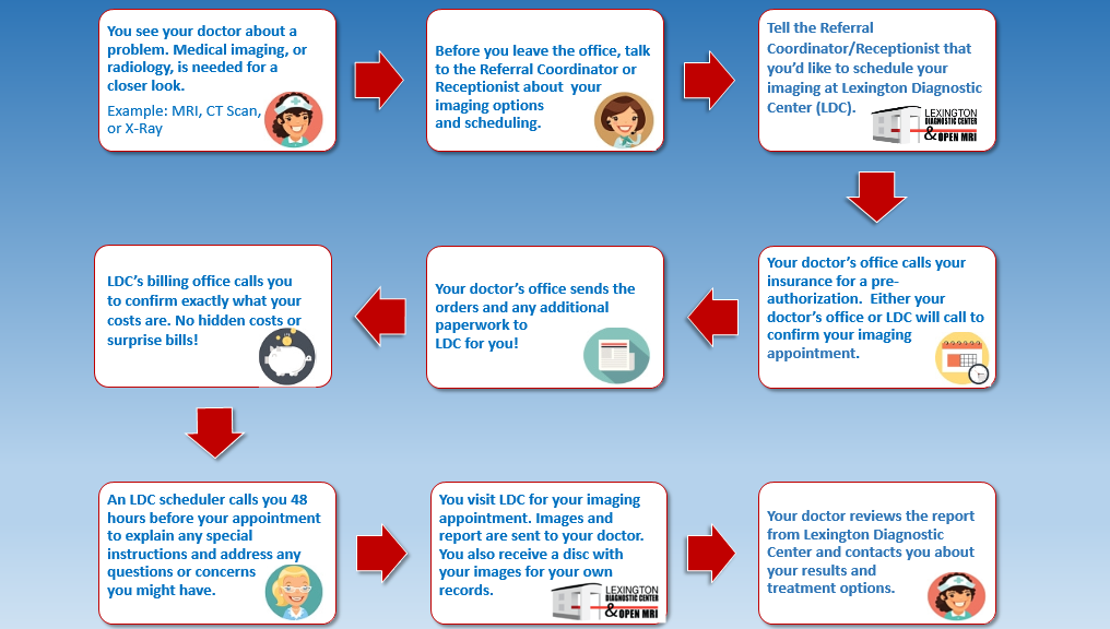
Often, diagnostic testing will be ordered: A CT scan, an MRI, or an ultrasound to diagnose and, on occasion, follow-up exams to evaluate treatment. Lexington Diagnostic Center and Open MRI specializes in diagnostic imaging ? and anxiety reduction. That?s because imaging exams at LDC are considerably less expensive than the hospital. And, LDC patients always know ? up front ? what testing is going to cost. There are no hidden fees or surprise bills that arrive weeks or months later.

Price transparency is one part of Lexington Diagnostic Center?s tradition of treating patients like family.

?We are such a family here at Lexington Diagnostic Center,? said radiologist Jason Harris, M.D., who joined the center in 2012. ?We have the best trained team, a strong commitment to quality, and a tremendous focus on doing the right thing for our patients,? he said.

His commitment to the LDC family is so strong, in fact, that he recently purchased the facility ? the only locally owned imaging facility in Lexington ?? from retiring founder George Privett, M.D.

Being a part of Lexington Diagnostic Center allows Dr. Harris to do what he loves most: Make a difference in peoples? lives. ?Every morning, when I come to work, before I start, I spend a few minutes thinking about what I am doing. With each study I read, I remind myself this study represents a person, somebody with a life and a family.

?I try to think of each one as a member of my family, as someone I know. Thinking this way motivates me to give it my absolute best each time,? he said.

The desire to give his best to his patients led Dr. Harris to study radiology at two of the best radiology programs in the United States. His radiology residency was completed at the University of Cincinnati, a world-renown center for neuroradiology. Following five years of residency at UC, Dr. Harris did a fellowship in musculoskeletal radiology at the Medical College of Virginia/Virginia Commonwealth University in Richmond, Va.

?Doing the fellowship required a lot of sacrifice,? Dr. Harris said, ?but I absolutely feel it was worth it. Having that additional training and experience has enhanced my practice immensely. It allows me to offer more and higher quality care to patients here at the Center.? The fellowship focused on diagnosing and treating conditions affecting the bones, joints, connective tissues, muscles and spine, including image-guided biopsies and joint injections.

Because of this additional training, Dr. Harris sometimes finds himself performing joint injections and other treatments. This direct interaction is something he enjoys. ?Every doctor in practice today woke up one morning and said to themselves, ?I want to be a doctor, I want to help people,?? he noted. That is, ultimately, what medicine is all about.

As Lexington Diagnostic Center?s new owner, Dr. Harris is committed to carrying on the center?s tradition of helping people by providing the highest level of quality and service, saving them money on their exams.

It?s an important point. Medical costs continue to rise and hospital-based services have grown even more expensive. As a result, free-standing, independent facilities like Lexington Diagnostic Center have a larger role to play in providing patients with affordable, high-quality services. LDC can save patients money because all they do is imaging. There is no hospital overhead ? laundry, meal services, ER team, administration.

Those additional expenses force hospitals to charge up to six times as much for the same exam. Because patients are often responsible for 20 to 50 percent of allowed charges, this often results in more cost for the patient.

?We don?t do everything a hospital does. We do imaging. And we do it well. And we save patients money,? Dr. Harris said.

Making Outstanding Care, Superior Quality Convenient and Affordable

Scan Plan Chart

When it is time for an imaging study ? CT scan, MRI, ultrasound ? many patients simply ?go with the flow? in the belief that there are no choices or that all of their choices are, essentially, the same.

Nothing could be further from the truth!
Lexington Diagnostic Center and Open MRI, located on Harrodsburg Road in Lexington, offers patients not only outstanding, personal care but superior quality. With full-time board-certified and fellowship-trained radiologists in the center daily; experienced, certified technical staff performing the exams; a laser-like focus on the patient and family; and state-of-the-art equipment, Lexington Diagnostic Center is able to deliver a much higher level of care than is available anywhere else.

?Diagnostic imaging is all we do,? said Davonna Saier, marketing manager for the center. ?This singular focus allows LDC to schedule patients quickly, conduct tests efficiently, provide outstanding quality and reporting and do it all at a cost significantly less than what patients might pay elsewhere.?

Patients are sometimes concerned that imaging studies performed outside a hospital setting won?t be readily available to their physicians, or cannot be compared to previous studies. ?That is absolutely not a concern,? Saier said. ?Lexington Diagnostic Center provides an advanced Picture Archiving and Communications System (PACS) which allows the referring physician to access our radiologists? reports and imaging studies from their own computers, or even from the surgery suite at the local hospitals,? Saier said.

Further, every patient who has imaging performed at LDC leaves the center that day with a disk containing all of the images captured during the visit. ?They can take the images directly to their doctor for review and, of course, the images are always archived here at the center for future reference if needed.?

As for comparing today?s images to those captured months or even years ago, Lexington Diagnostic Center is able to request those images from other medical facilities for comparison purposes ? continuity of care ? without issue.

?We have a courier who runs between all the medical facilities daily picking up imaging studies and returning them here, to the Center, where our radiologists use them to compare today?s studies with any studies performed previously,? Saier said.

There is no charge to the patient for this service ? and no hassle either. ?All they need to do is tell us where they had the study done, and when, and we?ll take care of the rest,? Saier said.

Patients also have access to all of their studies performed at Lexington Diagnostic through a secure internet connection, the Patient Portal. The portal allows patients to read and study the radiologist?s findings and print them out if desired. ?This allows patients to look up terms, think about the findings and follow up with the referring physician with questions or concerns,? Saier noted.

It is one advantage of having imaging done at LDC that many patients really appreciate, she added. Another benefit is how convenient and easy it is to have testing done at Lexington Diagnostic. With early evening and Saturday hours, patients never have to wait weeks to have a test scheduled. When you arrive at the center for testing, you can feel confident your test will be performed and you?ll be on your way quickly. There are no long waits caused by emergency cases bumping the schedule. Results are reported to the referring physician very quickly, always in less than 72 business hours, Saier noted.

One of the most important considerations for patients receiving care anywhere is cost. Patients at Lexington Diagnostic Center have peace of mind knowing exactly how much their imaging study will cost, and what their out-of-pocket costs will be. Most patients will save hundreds of dollars by choosing to have their MRIs, CTs and other tests performed at LDC instead of the local hospital.

?That?s because hospitals have to cover all of that overhead ? the cafeteria, laundry, parking garages ? it?s all rolled up into every patient?s bill. At Lexington Diagnostic Center, imaging is the only thing we do. We keep our overhead costs low and that keeps our prices low, too.?

Taking advantage of the Lexington Diagnostic Center difference is quite easy. All you have to do is speak up when your doctor orders an imaging procedure for you. ?Most patients don?t realize they have the right to decide where they?ll go for a test or procedure,? Saier said. When patients fail to express a preference, the doctor?s office will schedule the procedure where it is most convenient for them ? usually the hospital.
Sometimes patients feel uncomfortable telling physician they need find a less expensive alternative to the hospital, Saier noted. That is natural. But, she added, ?Your doctor should be just as concerned about your financial health as he or she is about your physical health.?

Price transparence is an important part of the Lexington Diagnostic Center patient care philosophy. ?It?s important for patients to know not only how much a test will cost, but what they will be responsible for in terms of co-pays, deductibles or co-insurance,? Saier said. LDC works with the patient?s insurer to determine the out-of-pocket costs before testing is performed. Patients are informed up front, usually at least 48 hours before their appointment. Payment is collected on the day of the test.

LDC accepts all insurance plans, including Medicare and Medicaid. ?We?re also an excellent choice for self-pay patients because testing at our facility is very affordable,? she noted. LDC is so affordable that several insurance companies will encourage enrollees to reschedule at the center. ?We?ve actually had them conference in with the patient to reschedule from a higher-priced facility to us,? Saier added.

New CT Scanner Brings New Capabilities to Lexington Diagnostic Center

LightSpeed with ASiR CT Machine

Lexington Diagnostic Center and Open MRI is pleased to announce the installation of a new 16-slice CT scanner to better serve the needs of its patients and the community.

The GE Lightspeed CT scanner uses x-rays and advanced computer software to produce three-dimensional, color-enhanced images of the body. The CT scanner produces multiple cross sections of the body part being examined, which allows the radiologist to study each ?slice? to look for abnormalities.

CT scans are especially useful in diagnosing diseases and conditions of hard tissues, like bones and cartilage, as well as the neck, chest, abdomen and pelvis. The new equipment allows Lexington Diagnostic Center to offer a new service ? CT angiography.

?CT angiography allows us to capture images of the blood vessels,? said Chad Blair, clinical manager at Lexington Diagnostic Center. ?CTA is useful in diagnosing conditions that affect the blood vessels that supply blood to the brain, lungs, abdomen, kidneys, the arms and legs,? he noted.

Conditions detected by CTA include narrowing of the arteries, or atherosclerosis; blockages; weaknesses in the arteries (aneurysms); tears in the vessels (dissection); blood clots in the lungs; buildup of fluid around the heart (pericarditis); and abnormal blood vessel patterns that could indicate an arteriovenous malformation (AVM) or tumor.

The new CT scanner offers quicker scan times, a larger bore to accommodate patients up to 400 pounds and high-resolution images, Blair noted. An important feature of the new CT is ASiR technology, which can reduce radiation exposure by as much as 40 percent.

In addition to CT angiography, the new equipment can:
? Low-Dose CT, a relative new procedure that has been approved by the FDA for early detection of lung abnormalities, including those that could indicate cancer.
? Virtual colonoscopy, which allows patients to undergo this important, life-saving testing without the anxiety and invasiveness of a regular colonoscopy

?We are very pleased to be able to offer a wide range of high-quality CT exams to the community at costs that are often considerably less,? Blair said.

Discover the Lexington Diagnostic Center and Open MRI quality and price difference. For more information about having your next imaging exam performed at LDC, give us a call at (859) 278-7226.

The mission of Lexington Diagnostic Center & Open MRI is to provide high quality compassionate medical imaging services at fair prices for all patients throughout the commonwealth.

LEXINGTON DIAGNOSTIC CENTER ANNOUNCES NEW OWNERSHIP

Dr. Jason Harris Lexington Diagnostic Center and Open MRIJason S. Harris, MD, MBA, has been named Medical Director and Owner of Lexington Diagnostic Center & Open MRI (d/b/a Neurodiagnostics, Inc.). George W. Privett, Jr., MD is retiring and has sold the business. He will stay on staff as a consultant for the next year.

Dr. Harris is a board-certified musculoskeletal fellowship-trained radiologist and has been with Lexington Diagnostic Center since 2012.

?I am honored by this opportunity to lead LDC, and pledge to continue our proud tradition of providing quality medical imaging care,? says Harris.

Dr. Privett started the independent medical imaging business in 1984. The center offers MRI, CT, X-Ray, ultrasound, nuclear medicine and DEXA scans.

?Lexington Diagnostic Center & Open MRI has always been known for quality compassionate care and fair prices. We are delighted to welcome Dr. Harris as our new Medical Director, and look forward to his leadership as we continue our mission serving patients throughout the Commonwealth,? says Marketing Director, Davonna Saier.

# # #

If you would like more information, please contact Davonna Saier at 859.278.7226 or email at d.saier@ldcmri.com.

Hosted By BT Web Group